Molecular biology relies on protein biotinylation as a crucial technique that facilitates the covalent bonding of biotin with target proteins. The technology proves essential for protein analysis and purification because it allows researchers to label and follow proteins in different experimental contexts. This article examines biotinylation’s mechanisms and applications while discussing its advantages and limitations and comparing it with other protein labeling methods to highlight biotinylation’s role in modern biological research.
First, let’s understand what biotin is. Vitamin B7, which is biotin, represents a water-soluble B-vitamin molecule that weighs 244.31 Da. The structure of biotin features a fused ring system that includes a ureido ring together with a sulfur-containing tetrahydrofuran ring connected to a valeric acid side chain.
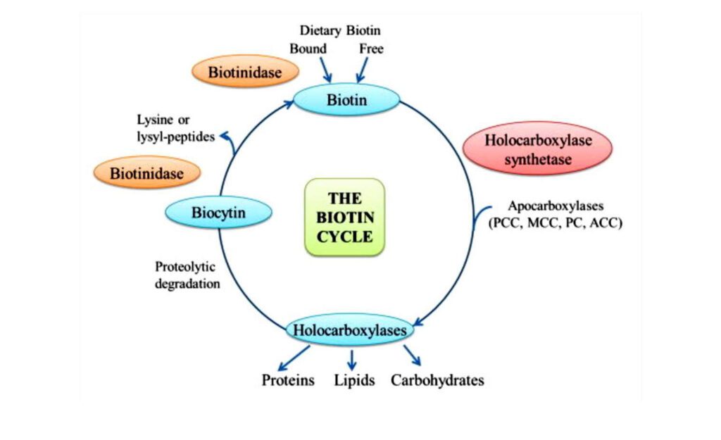
Biotin also serves as an essential cofactor for carboxylase enzymes, which participate in key metabolic pathways. The metabolic pathways impacted by these processes are fatty acid synthesis along with gluconeogenesis and amino acid metabolism. Biotin functions as a crucial agent for carbon dioxide group transfer during these processes, which makes it indispensable for both energy production and proper cellular functioning.
The protein biotinylation process involves the covalent attachment of biotin molecules to designated sites on target proteins. The biotin-avidin/streptavidin system forms the core of the technique by delivering exceptional specificity and stability essential for multiple biological applications.
Let us now examine how protein biotinylation functions as a biochemical process. Protein biotinylation reactions can be grouped into chemical biotinylation and enzyme biotinylation methods.
The chemical biotinylation process utilizes activated biotin derivatives like NHS esters to create covalent bonds with protein amino groups. Common reaction targets include primary amines (e.g., lysine ε-amino groups or N-terminal amino groups), thiol groups (e.g., cysteine), and carboxyl groups.
NHS-PEG4-Biotin reagents utilize a polyethylene glycol (PEG) spacer arm measuring 29 Å to connect biotin to proteins. The design increases aqueous solubility and minimizes steric interference, which allows for dependable amide bond creation under physiological pH conditions between pH 7 and 9.
Enzymatic biotinylation requires biotin ligases like BirA to attach biotin specifically to target proteins at peptide sequences, including the AviTag, which consists of 15 amino acids recognized by BirA. The enzymatic biotinylation method stands out because it attaches biotin precisely to proteins without altering their natural structures. The technique delivers superior labeling efficiency of over 95%, which makes it perfect for research that needs consistent biotin attachment.
Biotinylated Protein Isolation


