How to Get Better Images from Deep within Your Sample? - Labinsights

How to Get Better Images from Deep within Your Sample?

icon.highlightedarticle.dark Microscopes
Last modified: 21 October 2025
Brain_sagittal_4x_whole
Brain_sagittal_4x_whole | Photo: Analis sa/ nv
Article image of: How to Get Better Images from Deep within Your Sample?
ANALIS - EVIDENT VS200 Slide Scanner’s & SILA | Photo: Analis sa/nv
Article image of: How to Get Better Images from Deep within Your Sample?
ANALIS - EVIDENT VS200 Slide Scanner’s & SILA | Photo: Analis
Article image of: How to Get Better Images from Deep within Your Sample?
EVIDENT VS 200 - Brain_sagittal_4x_whole | Photo: Analis
Article image of: How to Get Better Images from Deep within Your Sample?
ANALIS - EVIDENT SLIDEVIEW VS200 for Life Science Research | Photo: Analis
Article image of: How to Get Better Images from Deep within Your Sample?
ANALIS - EVIDENT SLIDEVIEW VS200 for Digital Pathology | | Photo: Analis

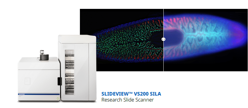
With the EVIDENT VS200 Slide Scanner & SILA Optical Sectioning Device

The Evident SLIDEVIEW VS200 is a universal whole slide imaging scanner.

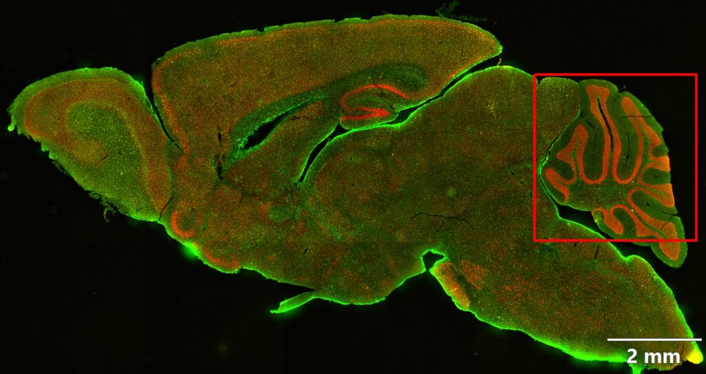
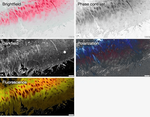
The VS200 system is designed to capture a wide variety of samples and stains with multiple magnifications, flexible slide sizes, and five versatile imaging modes. Outstanding image quality of entire slides empowers your lab to unlock the full potential of your samples.

Discover more details in your samples with five imaging modes—brightfield, polarization, fluorescence, darkfield, and phase contrast in one system—and the ability to combine multiple techniques in a single scan.

Reveal More Sample Details with 5 Flexible Imaging Modes

Discover more details in your samples with five imaging modes—brightfield, polarization, fluorescence, darkfield, and phase contrast in one system—and the ability to combine multiple techniques in a single scan.

  • Application field: Life Science Research and Digital Pathology

SILA Optical Sectioning Device

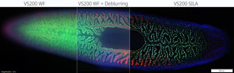
Easy to install, the Speckle Illumination Acquisition (SILA) device is compact and easily attaches to the VS200 system’s fluorescent illuminator.

Speckles are used to obtain high-contrast images, removing out-of-focus light to deliver sharp images, especially from thick samples. The image is computed during the scan, and there’s no need for post-processing, so the device has a minimal impact on acquisition speed.

You are welcome to contact our experts.

EXPLORE MORE icon.arrow--dark

Written by

Logo of:How to Get Better Images from Deep within Your Sample?

Analis sa/nv

Analis, distributeur & fabrikant van instrumenten en meubilair voor laboratoria. Meer dan een vertrouwenslabel, UW KWALITEITSLABEL Read more