Brucellosis is a zoonotic infectious disease caused by Brucella, and it is one of the most widespread zoonotic diseases in the world, which brings great losses to the animal husbandry industry. Here, we will outline several aspects about the pathogen, living environment, identification, and diagnostic procedures of Brucella, hoping to popularize knowledge about brucellosis. Characteristics of Brucella pathogens Brucella colonies are small, immobile, ball-shaped, and are Gram-negative bacteria. Acid-fast staining was positive due to inability to destain by a modified acid-fast staining technique with 0.5% acetic acid. Acid-fast staining of Brucella in body fluids or tissue smears shows characteristic clusters of Rhodococcus. Due to the high degree of genetic similarity of Brucella, it has been suggested to be classified as Brucella malta. However, this decision changed in 2005, when 6 Brucella species were re-identified. In addition, some new species, including Brucella from marine mammals and voles, are divided into Brucella abortus, Brucella malta, Brucella suis, and Brucella ovine according to their serological characteristics. , Brucella canis, Brucella sarin, Brucella dolphins, Brucella seals.
Brucella Living Environment
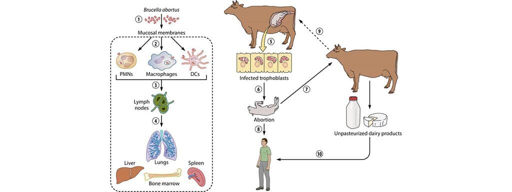
Brucella is usually present in the reproductive organs of sexually mature male and female animals, and each species of Brucella infects specific animal species. Infected animals often continue to infect other animals as a source of infection. Bacteria excreted by infected animals can survive for several months in moist environments. It is usually spread through direct contact with infected animals and abortion-related bodily fluids and tissues.
Identification of Brucella
Brucella can be identified by colony morphology, biochemical experiments, specific culture requirements, and growth inhibition of dyes. In addition, agglutination with monospecific serum, susceptibility to bacteriophage, and molecular biology methods are also used for bacterial species identification. Colonies of B. abortus, B. malta, and B. suis were small and smooth at the initial stage of isolation, and became glossy, bluish, and translucent colonies after 3 to 5 days. Colonies became opaque with increasing incubation time. In contrast, the colonies of B. ovine and B. canis were rough, dull, yellow, opaque, and brittle. Brucella is not hemolytic on blood agar medium.
Diagnostic Procedures for Brucella
The diagnosis of Brucella mainly relies on serological testing and bacterial isolation and identification. Samples should be transported and handled with great care and in a safety cabinet. Samples for laboratory testing must be associated with specific clinical symptoms. Examination of specimens with MZM-stained smears, especially placental villus lobules, fetal true stomach contents, and uterine secretions, often shows characteristic MZN-positive cocci. In samples containing cells, these bacteria may bleed in clusters. Samples can be tested by PCR. The advantage is that it is highly sensitive and can be used to detect small amounts of samples and microorganisms. Nutrient-rich media are commonly used for isolation. The medium can be stored for 5 days at 37°C in 5% to 10% carbon dioxide. Although carbon dioxide is a special substance required by individual bacterial species, most Brucella species are carbon dioxide-loving. In addition, serological testing can be used for international trade and to identify infected flocks and individual animals. Antigens from Brucella and some other Gram-negative bacteria, such as Yersinia enterocolitica serotype O:9, cross-react in agglutination assays.
